Cingulate Gyrus: Vascular Supply, Distributions of the Anterior and Posterior Cerebral Arteries
The cingulate gyrus is not supplied by an independent arterial system.
Rather, branches of the anterior cerebral artery supply most of cingulate cortex, adjacent frontal and parietal cortices, a few brainstem structures, and most of the corpus callosum. One consequence of this organization is that infarcts of even a single branch of the pericallosal system can lead to damage beyond cingulate cortex and produces complex neurological symptoms.
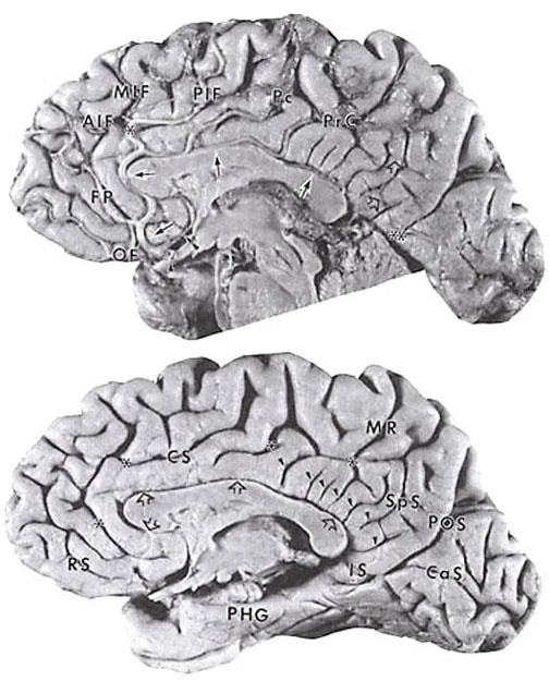
The anterior cerebral artery is a branch of the internal carotid artery, and it extends to the anterior communicating artery that is indicated with a single arrowhead in the Figure. The precallosal artery (double arrowhead in figure) is emitted from the initial part of the pericallosal artery and distributes to the rostrum of the corpus callosum (Perlmutter and Rhoton, 1978). The pericallosal artery is formed at the point where the anterior communicating artery crosses the midline. The pericallosal artery can emit all subsequent cortical and callosal branches. In most cases, however, there is a callosomarginal branch of the pericallosal artery (Perlmutter and Rhoton, 1978). When present, the callosomarginal artery is the largest branch of the pericallosal artery, and it runs into the cingulate sulcus. The origin can vary from rostral to the anterior communicating artery to just dorsal to the genu of the corpus callosum. The sharp angle at which the callosomarginal artery is emitted at the rostrum predisposes it to one of the highest incidences of aneurysms in the pericallosal system (Morris and Peck, 1955).
Medial surface of a 62-year-old, neurologically intact male.
Top: The pericallosal artery (arrows) begins at the junction of the anterior communicating artery (arrowhead) and its first branch is the precallosal artery (double arrowhead). The cortical branches include the common trunk of the orbitofrontal (OF) and frontopolar (FP) arteries, the paracentral (Pc) and precuneal (PrC) branches and the terminal branch of the pericallosal artery is at the far right arrow. The callosomarginal artery (asterisk) emits the anterior, middle, and posterior inferior frontal (AIF, MIF, PIF, respectively) arteries. Small parietooccipital arteries (open arrows) anastomose with the pericallosal artery and the medial branch of the posterior cerebral artery (double asterisk).
Bottom: The arteries and pia mater were removed to show the calcarine sulcus (CaS), callosal sulcus (open arrows), cingulate sulcus (CS) and asterisks, isthmus of the cingulate gyrus (IS) which is also termed the caudomedial subregion, marginal ramus of the CS (MR), parahippocampal gyrus (PHG), parietooccipital sulcus (POS), posterior cingulate dimples (arrowheads), rostral sulcus (RS) and splenial sulcus (SpS).
The pericallosal artery emits as number of short and long branches, the latter of which were clearly photographed by Perlmutter and Rhoton (1978). There are eight major cerebral branches of the pericallosal artery. When the callosomarginal artery is present, it most frequently emits the middle internal frontal artery. Marino (1976) described the branches of the anterior cerebral artery in autopsy material with vascular injections, and this article and that by Perlmutter and Rhoton (1978) should be consuted for the details of thedistribution for each branch.
The first and second cortical branches of the pericallosal artery are the orbitofrontal artery, which supplies the gyrus rectus and adjacent cingulate cortex, and the frontopolar artery, which distributes to the frontal pole. In the specimen shown in the Figure, there is a common trunk for these two arteries. The anterior, middle, and posterior inferior frontal arteries supply progressively more caudal parts of the cingulate gyrus dorsal to the corpus callosum. These arteries can arise as individual branches of the pericallosal artery or, as in the specimen in the Figure, the callosomarginal artery can emit all three inferior frontal arteries. The internal parietal, or paracentral, artery distributes to cortex of the paracentral lobule and the cingulate gyrus ventral to it, and to precuneal, or superior parietal, artery supplies precuneal and posterior cingulate cortices. Finally, the parietooccipital, or inferiorparietal, artery can be a branch of the pericallosal artery and can supply cingulate cortex above the splenium of the corpus callosum, or the terminal branches of the pericallosal artery may anastomose with the precuneal artery and parietooccipital branch of the posterior cerebral artery to supply this caudal part of the cingulate gyrus.
The most caudal extension of cingulate cortex is termed the isthmus or caudomedial region and it is supplied by small branches of the medial branch of the posterior cerebral artery (Zeal and Rhoton, 1978; Waddington, 1984). This artery can also distribute to cuneal and precuneal cortex and supplies parts of the midbrain and thalamus (Zeal and Rhoton, 1978). A terminal branch of the parieto-occipital artery is the splenial artery. This artery can distribute to the splenium of the corpus callosm and frequently anastomoses with the terminal branches of the pericallosal artery.
References
Marino R (1976) The anterior cerebral artery: I. Anatamo-readiological study of its cortical teritories. Surg Neurol 5:81-87.
Morris AA, Peck CM (1955) Roentgenographic study of the variations in the normal anterior cerebral artery. Am J Roetgenol Radium Ther Nuc Med 74:818-826.
Perlmutter D, Rhoton AL (1976) Microsurgical anatomy of anterior cerebral-anterior communicating-recurrent artery complex. Surg Forum 27:464-465.
Perlmutter D, Rhoton AL (1978) Microsurgical anatomy of the distal anterior cerebral artery. J Neurosurg 49:204-228.
Waddington MM (1984) Atlas of Human Intracranial Anatomy, Rutland, VT: Academy Books.
Zeal AA, Rhoton AL (1978) Microsurgical anatomy of the posterior cerebral artery. J Neurosurg 48:534-559.

