About CNSI
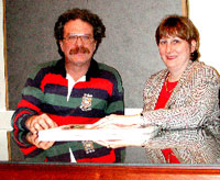
History. Cingulum NeuroSciences Institute (CNSI) is a nonprofit and tax-exempt, 509(a)(2) corporation (EIN 56-2020907) formed in 1997 by Dr. Brent A. and Leslie J. Vogt (pictured below). The name is a registered Service Mark with the US Trademark and Patent Office and refers to the cingulum bundle which is a complex of mostly myelinated axons that underlies cingulate cortex. In 2000 we moved from North Carolina to Upstate New York where we are a registered charity (#41-63-33). CNSI is built on the past 50 years of expertise derived from the studies of Dr. Vogt into the structure, connections, functions, and pathologies of the cingulate gyrus in rodent, lagomorph (rabbit), and primate (monkey & human) brains (see Dr. Vogt’s CV).
Mission. We are neuroscientific entrepreneurs that seek to maintain a high scientific standard, morphological and imaging data bases, and research facilities to enhance our ability to add value as consultants and collaborators in the cingulate neurosciences. This includes imaging craftsmanship/brain anatomy, translation of cingulate animal research to human imaging and diseases, support of cingulate research with information, and resources including books, meetings and this web site. We collaborate with many neuroscientists in the US and throughout the world including the U.K., Germany, Taiwan and Singapore. (See pictures of our collaborators.) We actively support the 8-subregion cingulate cytoarchitectural map and continue to drive its evolution to incorporate functional and disease states.
Harsh Physical Child Abuse & Rape. Survivors of these events suffer psychopathologies both during adolescence and as adults; see the section titled “Psychiatric Damage Evoked by Harsh Child Abuse” & “Epidemiology of Adult-Onset Psychopathology Following Child Maltreatment” that explain how child abuse damages the brain including cingulate cortex. We have developed an animal model of child abuse disorder; see Vogt, Vogt, Sikes (2017; article #66 in Library). Defining the brain changes and searching for a cure is our primary, disease mission.
CNSI brings hope for survivors through brain research.
Expertise
We have two primary goals.
We have two primary goals.
- To understand the structure/function organization of each cingulate subregion in health and disease. This effort also focuses on the essential functions of individual areas. As such we have a uniquely cingulocentric perspective based in rigorous anatomy that helps to interpret a range of anatomical/structural and functional imaging data in rodents and primates, including humans. Thus, we assist in the design and interpretation of structural and functional imaging studies as well as in training postdoctoral fellows or early faculty with an evolving interest in cingulate cortex. We collaborate in joint grant applications and other activities that have a significant focus on cingulate cortex. Our facilities support these activities with a full histological preparation and analysis suite including five microscopic imaging systems and a library of histological cingulate tissues from five species including numerous human diseases.
- To understand and eventually treat the consequences of child abuse and rape by using an animal model of these experiences. This research is in its earliest stages as we now need to understand how these events evolve into adult-onset psychopathology. The ultimate goal is to block these changes so that individuals that are vulnerable to abuse can live their life free of the agony of such treatment.
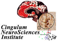
The company logo is a coregistration of structural magnetic resonance images (gray) of Dr. Vogt's brain and positron emission tomography images of binding of the opiate receptor antagonist diprenorphine (Library Item #95). Heaviest binding is in red in anterior and midcingulate cortices. There is also an image of neurons expressing intermediate neurofilament proteins in the caudal cingulate motor area that project to the spinal cord (Item #9). The third image is a three-dimensional, eigenvector projection of values of neuron densities in posterior cingulate area 23 for five statistical subgroups of Alzheimer's disease (Item #90 in the CNSI Neuroscience Library).


